Research Peptides
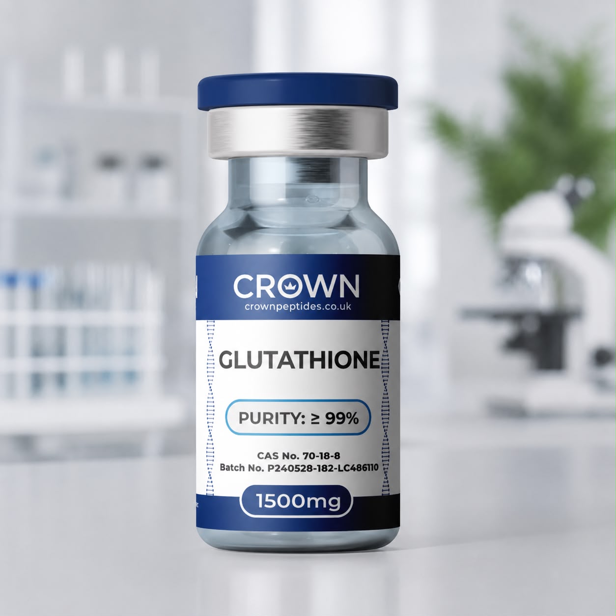
A high-purity research compound studied in antioxidant and cellular-protection research. Available in 1500mg, with every batch independently tested and dispatched from the UK.
Glutathione peptide UK is Crown Peptides' naturally occurring tripeptide research compound, available in 1500mg strength for laboratory research.
Supplied for laboratory research purposes only — not for human consumption or for medical, veterinary or diagnostic use.
Glutathione is a tripeptide composed of glutamate, cysteine and glycine. It is one of the most important intracellular redox molecules and is widely used in biochemical research examining oxidative stress, thiol chemistry, cellular antioxidant systems and glutathione-dependent enzymes.
Also known as: GSH, L-glutathione, reduced glutathione.
Research uses glutathione to study the balance between reduced glutathione and its oxidised form, glutathione disulfide — a redox couple that provides a direct read-out of cellular oxidative state. Its protective role is well documented: in cultured human skin fibroblasts, glutathione protected the cells against the damaging effects of UVB and UVA light, with protection in the UVB range approaching that of the cells’ own DNA repair (Learn more). Glutathione is also central to glutathione peroxidase and glutathione transferase pathways, xenobiotic metabolism and broader research into mitochondrial and cellular redox homeostasis, alongside related redox-research compounds such as Methylene Blue.
Most peptides are strung together on ribosomes from a genetic template. Glutathione is not. Cells assemble it in two ATP-driven steps: glutamate-cysteine ligase first joins glutamate to cysteine through glutamate’s side-chain carboxyl group, and glutathione synthetase then adds glycine. That first linkage, the gamma-glutamyl bond, is why the molecule is written γ-Glu-Cys-Gly, and it makes glutathione resistant to the ordinary peptidases that chew through conventional peptides inside the cell. Only a specialised enzyme, gamma-glutamyl transpeptidase, which sits on the outer surface of many cells, can cleave it.
The ligase step sets the pace. It depends on cysteine supply and is slowed by glutathione itself through feedback inhibition, which gives cells a built-in thermostat for their glutathione pool. For laboratories, this pathway is a gift: buthionine sulfoximine, a selective inhibitor of the ligase described by Griffith and Meister in the late 1970s, lets researchers drain glutathione from cultured cells in a controlled way, and purified glutathione is the natural reference standard against which that depletion is measured.
When glutathione donates electrons, two molecules join through a disulfide bridge to form glutathione disulfide, GSSG. Glutathione reductase reverses the reaction using NADPH, so the ratio of the two forms acts like a live gauge of how hard a cell is working to stay reduced. In the cytosol of healthy cells the reduced form heavily outnumbers the oxidised form, and total glutathione typically sits in the low millimolar range, far above most other small thiols.
That gauge reads differently from one compartment to the next. The endoplasmic reticulum, where new proteins form their disulfide bonds, keeps a markedly more oxidising glutathione balance than the surrounding cytosol, and published ER ratios fall to single figures. Researchers track these differences with two main tools: the classic Tietze enzymatic recycling assay, which uses glutathione reductase and Ellman’s reagent (DTNB) to quantify total and oxidised glutathione in extracts, and genetically encoded sensors such as Grx1-roGFP2, which report the glutathione redox potential in living cells in real time. Both rely on well-characterised glutathione for calibration.
One of the most active areas in redox biology over the past decade began with a puzzle: certain compounds killed cancer cell lines by a route that looked nothing like apoptosis. In 2012, Dixon and colleagues named this iron-dependent, lipid-peroxide-driven process ferroptosis, and glutathione turned out to sit right at its centre.
The key enzyme is glutathione peroxidase 4 (GPX4), which uses glutathione to convert dangerous lipid hydroperoxides in membranes into harmless lipid alcohols. Starve GPX4 of glutathione and peroxidised lipids accumulate until the membrane fails. The small molecule erastin does exactly that by blocking system xc−, the transporter that imports cystine for glutathione synthesis, while RSL3 inhibits GPX4 directly. Measuring how glutathione levels fall, and whether adding glutathione back changes the outcome, has become a standard experimental design in ferroptosis research across cancer, neuronal and kidney cell models.
Beyond being a molecule to measure, glutathione is a reagent that cell biologists add, remove and rescue. Depletion experiments with buthionine sulfoximine are often paired with add-back conditions to show that an effect truly depends on the glutathione pool rather than an off-target action of the inhibitor.
There are practical twists. In culture medium, the free thiol slowly oxidises, especially in the presence of trace metals and at neutral to slightly alkaline pH, so the reduced form does not stay reduced indefinitely. And many cell types take up little intact glutathione from outside: gamma-glutamyl transpeptidase on the cell surface breaks it into its amino acids, which are then reabsorbed and used to rebuild glutathione inside. This is why membrane-permeable derivatives such as glutathione monoethyl ester were developed in Alton Meister’s laboratory, and why careful study designs compare native glutathione, its esters and cysteine precursors side by side.
Compound-level reference data. Batch-specific results appear on the Certificate of Analysis.
Work out concentration with the peptide calculatorCrown Peptides Glutathione is supplied with batch-specific quality documentation appropriate to the material. Analytical methods vary by compound and formulation, so the Certificate of Analysis should be used as the definitive source for identity, assay, purity and relevant impurity information.
Researchers working with Glutathione may also be interested in Methylene Blue, MOTS-c, SS-31, NAD+ and 5-Amino-1MQ — other compounds studied in cellular redox, oxidative-stress and mitochondrial research.
Glutathione is the tripeptide γ-glutamyl-cysteinyl-glycine (C10H17N3O6S, about 307.32 g/mol), the most abundant non-protein thiol in animal cells. Crown Peptides supplies the reduced form, often labelled GSH, L-glutathione or reduced glutathione, in 1500mg vials for redox, thiol and enzyme research.
It is, with a twist. Glutamate is attached through the carboxyl group on its side chain rather than the one beside its amino group, forming a gamma-peptide bond. The cysteine-glycine link is entirely conventional. That gamma linkage is what shields glutathione from most intracellular peptidases.
For eight years, it was. Frederick Gowland Hopkins coined the name glutathione in 1921 for what he took to be a two-residue peptide, then corrected it to a tripeptide in 1929, the same year Edward Kendall’s group crystallised it. The molecule had been glimpsed earlier still: in 1888 the French scientist J. de Rey-Pailhade described a sulphur-reducing substance in yeast that he called philothion. Chemical syntheses followed in the mid-1930s.
Two enzymes do the work. Glutamate-cysteine ligase forms γ-glutamylcysteine, and glutathione synthetase adds the glycine, each step consuming ATP. The genes encode these enzymes rather than the tripeptide itself, so glutathione levels track enzyme activity and cysteine availability.
GSSG is glutathione disulfide: two glutathione molecules joined through their cysteine sulphur atoms after giving up electrons. GSH is the reduced, single-molecule form with a free thiol group. Our product is supplied as GSH, and the balance between the two is what redox researchers measure.
Its thiol (–SH) group is what reacts. It can donate electrons to peroxides via peroxidase enzymes, attack electrophilic compounds during conjugation, form mixed disulfides with proteins and bind metal ions. Glutamate and glycine mainly govern how enzymes recognise and handle the molecule.
Glutathione transferases attach it to electrophilic and foreign compounds, the first step of the mercapturic acid pathway in detoxification research. Glutaredoxins use it to reduce protein disulfides, glyoxalase I needs it to process methylglyoxal, and glutathione reductase recycles GSSG back to GSH using NADPH.
It indicates how reducing a cell or compartment is at that moment. A falling ratio suggests oxidative load is outpacing recycling by glutathione reductase. Because the ER runs far more oxidised than the cytosol, researchers increasingly measure the ratio compartment by compartment rather than as one whole-cell number.
GPX4, the enzyme that protects membranes from lipid peroxidation, cannot work without it. Compounds such as erastin trigger ferroptosis in cell lines by cutting off the cystine needed to make glutathione, so glutathione measurements and add-back experiments are central to showing that a cell death is ferroptotic.
It is the reversible attachment of glutathione to a cysteine on a protein, forming a mixed disulfide. It can shield vulnerable cysteines from irreversible oxidation and switch enzyme activity on or off, and glutaredoxins remove it again. Purified glutathione is used to induce and study the modification in vitro.
Through NADPH. Glutathione reductase depends on NADPH to regenerate GSH, and cellular NADP+ is made from NAD+, so the two pools are linked. Methylene Blue is a redox-cycling dye that acts on glutathione reductase in enzyme and parasite studies, which makes it a natural companion when probing how the glutathione system copes with redox pressure.
Cells hold glutathione at millimolar levels, so standards, buffers and culture supplements are made at concentrations far higher than signalling peptides need. At roughly 4.9 millimoles of glutathione per 1500mg vial, one vial supports plenty of assay calibration or medium preparation. Labs running depletion and rescue series over many weeks can take five or ten vials at once, keeping every preparation on the same batch.
Dissolved oxygen oxidises the free thiol, and trace copper or iron speed that reaction up, as does a neutral or alkaline pH. Many protocols prepare solutions fresh, use degassed water or add a metal chelator such as EDTA, and keep working stocks cold to hold the reduced form steady.
Reduced glutathione appears as its protonated ion near m/z 308. A second ion near m/z 613 is the disulfide, formed when two molecules oxidise together in solution or in the ion source. Seeing how much 613 sits alongside 308 is a quick check on how much of a sample has oxidised, and it confirms the free cysteine thiol is present in the structure.
Often only partly. In many cell types, gamma-glutamyl transpeptidase on the outer membrane breaks extracellular glutathione into amino acids that are reabsorbed and reassembled inside. Researchers therefore compare native glutathione with its monoethyl ester or with cysteine precursors to separate effects outside the cell from changes in the internal pool.
Every batch is published openly — identity, purity and batch number, with no account needed.
Supplied for laboratory research purposes only — not for human consumption or for medical, veterinary or diagnostic use.
Each batch undergoes internal analytical verification using chromatographic and spectrometric methods prior to lyophilisation.
• HPLC Analysis
• Mass Spectrometry Confirmation
• Batch Traceability
By purchasing this product you confirm that you are a qualified researcher and that this compound will be used exclusively for laboratory research purposes.
This product is not intended for diagnostic or therapeutic use.
Any human or medical use is strictly prohibited.
Store in a cool, dry, secure and sterile environment away from any unauthorised persons or children.
Disclaimer: All products are sold strictly for laboratory research purposes only. Not for human or veterinary use, consumption, therapeutic, or diagnostic application. None of our products are medicines, and none are presented as such. By purchasing, you confirm you are a qualified professional and legally permitted to handle these materials in compliance with all applicable laws and regulations. Misuse, resale for unauthorised purposes, or unlawful application is strictly prohibited. Crown Peptides UK disclaims all liability for improper use, handling, or regulatory non-compliance.