Research Peptides
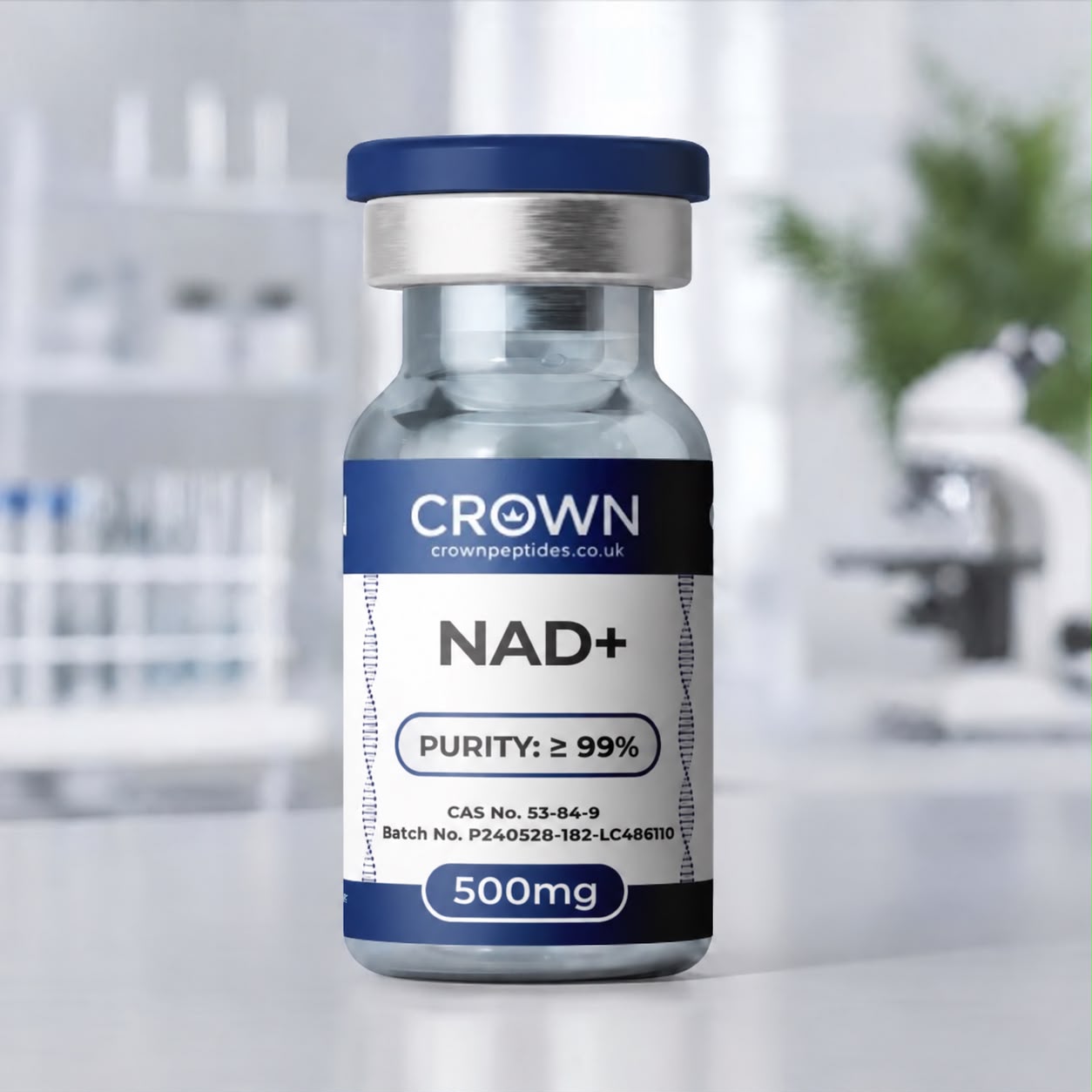
A high-purity research compound studied in cellular-energy and ageing research. Available in 500mg and 1000mg, with every batch independently tested and dispatched from the UK.
NAD+ compound UK is Crown Peptides' nicotinamide adenine dinucleotide research cofactor, supplied in 500mg and 1000mg vials for laboratory research into redox chemistry, cellular energy metabolism and sirtuin-dependent signalling.
Supplied for laboratory research purposes only — not for human consumption or for medical, veterinary or diagnostic use.
NAD+ is an essential dinucleotide cofactor present in virtually all living cells. It participates directly in oxidation-reduction reactions and also serves as a substrate for enzymes including sirtuins, PARPs and CD38, making it central to research on energy metabolism, DNA-damage signalling and cellular regulation.
Also known as: NAD, NAD plus, nicotinamide adenine dinucleotide, β-NAD.
NAD+ research spans mitochondrial metabolism, glycolysis, redox state, sirtuin-dependent signalling, poly-ADP-ribose polymerase activity and cellular stress responses, with the NAD+/NADH ratio widely used as a biochemical marker of cellular redox balance (Learn more). Research applications include energy metabolism, DNA-damage-response signalling and gene regulation, alongside related metabolic and ageing-research compounds such as MOTS-c and SS-31. Because NAD+ is chemically reactive and hygroscopic, batch documentation should be used to confirm the identity and condition of the specific material supplied.
The NAD+ story begins in London in 1906, when Arthur Harden and William Young were studying how cell-free yeast juice ferments sugar. Passing the juice through a membrane split it into two fractions: a large, heat-sensitive protein portion and a small portion that survived boiling. Neither could ferment glucose on its own, yet mixing them back together restored activity. The small, heat-stable factor was named cozymase.
Working out what cozymase actually was took another two decades. In Stockholm, Hans von Euler-Chelpin purified it and showed it to be a nucleotide containing adenine, nicotinamide, two ribose sugars and two phosphates, and he shared the 1929 Nobel Prize in Chemistry with Harden for their fermentation work. In the mid-1930s Otto Warburg’s laboratory in Berlin traced the chemistry to the nicotinamide ring, the part that picks up hydrogen and hands it on, and identified a phosphorylated sister molecule now called NADP+. Older papers call it diphosphopyridine nucleotide (DPN) or coenzyme I.
In redox chemistry NAD+ behaves as a shuttle rather than a fuel. Its nicotinamide ring accepts a hydride ion, two electrons and a proton delivered together, to become NADH; when NADH passes that hydride to another enzyme, NAD+ is regenerated intact. Glycolysis relies on this at the glyceraldehyde-3-phosphate dehydrogenase step, the citric acid cycle loads electrons onto NAD+ at three separate dehydrogenases, and complex I of the respiratory chain takes them off again to drive oxygen consumption.
The balance differs sharply from one compartment to another. Classic measurements in freeze-clamped rat liver by Williamson, Lund and Krebs put the free cytoplasmic NAD+/NADH ratio at about 725 in well-fed animals, while the mitochondrial matrix came out in single figures, a far more reduced environment. NADH cannot cross the inner mitochondrial membrane directly, so shuttle systems such as malate–aspartate carry reducing equivalents across instead. The reduced form also gives researchers a convenient handle: NADH absorbs light at 340 nm and fluoresces blue, while NAD+ does neither, so countless dehydrogenase assays and tissue autofluorescence studies simply follow that signal.
A second side of NAD+ biology appeared when researchers found enzymes that break the molecule apart instead of borrowing its electrons. In 2000, Shin-ichiro Imai, Leonard Guarente and colleagues reported in Nature that yeast Sir2, a gene-silencing protein linked to yeast lifespan, is an NAD+-dependent histone deacetylase. Mammals carry seven relatives, SIRT1 to SIRT7, spread across the nucleus, cytoplasm and mitochondria. Each strips an acetyl or other acyl group from a lysine on its target protein and cleaves NAD+ in the process, releasing nicotinamide, so sirtuin activity is tied directly to how much NAD+ a cell can spare. Free nicotinamide in turn inhibits sirtuins, a feedback loop experimenters have to allow for.
CD38 is a far hungrier consumer. This membrane enzyme, familiar as an immune-cell marker, hydrolyses NAD+ to nicotinamide and ADP-ribose and also makes cyclic ADP-ribose, a calcium-releasing messenger. In a 2016 Cell Metabolism study from the Mayo Clinic, Camacho-Pereira and colleagues found CD38 levels climbed with age in mouse tissues, while mice lacking CD38 held on to NAD+ and mitochondrial function as they aged, through a mechanism that depended on the mitochondrial sirtuin SIRT3.
Because sirtuins, PARPs and CD38 keep turning NAD+ into nicotinamide, cells need a quick way to rebuild it. In mammals the main route is salvage. Nicotinamide phosphoribosyltransferase (NAMPT) adds a phosphoribose group to nicotinamide to make nicotinamide mononucleotide (NMN), the rate-limiting step, and NMN adenylyltransferases then attach the adenosine half to complete the cofactor. Three versions, NMNAT1 to NMNAT3, work in the nucleus, the Golgi and cytoplasm, and mitochondria, so each pool is topped up locally. The selective NAMPT inhibitor FK866 drains NAD+ from cultured cells, making it a standard tool for testing which effects truly depend on the cofactor. Cells can also start from nicotinic acid, or build NAD+ from scratch out of tryptophan. Nicotinamide that leaves the loop by methylation is a separate story, told on our 5-Amino-1MQ page.
In 2004 Pawel Bieganowski and Charles Brenner described another entry point: nicotinamide riboside (NR), phosphorylated by NR kinases in a route conserved from yeast to humans, and they identified NR as a nutrient in milk. NR and NMN are now the two precursors most often seen in cell and animal studies. More recently, the 2020 identification of SLC25A51 as the carrier that imports NAD+ into mammalian mitochondria settled a long-running question about how the matrix pool is supplied.
Compound-level reference data. Batch-specific results appear on the Certificate of Analysis.
Work out concentration with the peptide calculatorCrown Peptides NAD+ is supplied with batch-specific quality documentation appropriate to the material. Analytical methods vary by compound and formulation, so the Certificate of Analysis should be used as the definitive source for identity, assay, purity and relevant impurity information.
Researchers working with NAD+ may also be interested in MOTS-c, SS-31, Methylene Blue, 5-Amino-1MQ and L-Carnitine (LC216) — other compounds studied in mitochondrial, redox and cellular-energy research.
Nicotinamide adenine dinucleotide is two nucleotides joined back to back, nicotinamide mononucleotide and adenosine monophosphate, linked through a pair of phosphates. It contains no amino acids, so it is a cofactor rather than a peptide (CAS 53-84-9, molecular weight about 663.43). Crown Peptides supplies it in 500mg and 1000mg vials, and it is also listed as NAD, NAD plus or β-NAD.
Two electrons and a proton. NAD+ is the oxidised, electron-accepting form; NADH is the reduced form, carrying a hydride on its nicotinamide ring ready to hand to another enzyme. Because the two interconvert constantly, most experiments track the ratio between them rather than either one alone.
The plus marks a positive charge on the nitrogen of the nicotinamide ring, which disappears when the ring takes on a hydride to become NADH. It describes the ring only: the two phosphates are negatively charged, so at neutral pH the whole molecule actually carries a net negative charge.
The geometry of the bond joining nicotinamide to its ribose sugar. Living cells make and use the β-anomer; the alternative α arrangement is a known isomer that most dehydrogenases do not accept, so specifying β-NAD confirms the biologically relevant form.
The nicotinamide ring is the vitamin. Nicotinic acid and nicotinamide, together called niacin or vitamin B3, are the raw materials cells turn into NAD+. The link was made in 1937, when Conrad Elvehjem’s group at the University of Wisconsin showed that nicotinic acid cured black tongue in dogs, the canine counterpart of pellagra.
By one extra phosphate on the adenosine ribose, added by NAD kinase. That small tag lets enzymes tell them apart, and cells keep them in opposite states: NAD+ mostly oxidised for energy metabolism, NADP mostly reduced as NADPH for biosynthesis and antioxidant defence. Every NADP molecule starts life as NAD+.
SARM1 is an enzyme in nerve fibres that, once triggered by injury, rapidly destroys NAD+ through its TIR domain, driving the axon self-destruct programme called Wallerian degeneration. It also explained an older puzzle: the WldS mouse, whose severed axons survive for weeks, carries a fusion protein containing the NAD+-making enzyme NMNAT1. Sustaining axonal NAD+ production, or switching off SARM1, delays breakdown in rodent and fly models.
PARP1 binds broken DNA and strings ADP-ribose units taken from NAD+ into long, branched chains that recruit repair proteins, releasing a nicotinamide with every unit. Heavy damage can drain a cell’s NAD+ and, with it, ATP production. PARP inhibitors such as olaparib are licensed cancer medicines, and in the lab they reveal how much NAD+ turnover the PARP family accounts for.
Much of it comes down to what happens outside the cell. Surface enzymes such as CD38 and CD73 break down NAD+ and NMN added to culture medium, and isotope-labelling work published in 2016 found that cells convert extracellular NMN to NR before uptake. NAD+ in the medium can still feed cells through those products: in a 2013 study, low micromolar NAD+ rescued tumour cells from FK866, a rescue that relied on CD73.
Extraction has to be split. Acid preserves NAD+ while destroying NADH, and alkali does the reverse, so labs often prepare two samples from the same tissue. Extracts then go to enzymatic cycling assays, which amplify tiny amounts into a colour or fluorescence signal, or to LC-MS, which quantifies NAD+, NADH and their precursors in one run. In living cells, genetically encoded sensors report the ratio in individual compartments in real time.
Through complex I, where NADH normally unloads its electrons. Methylene Blue can take electrons from NADH and deliver them to cytochrome c, opening a detour around blocked complexes in isolated mitochondria and cell studies, so pairing the two shows how NADH supply responds when that detour is open.
The bond between nicotinamide and ribose is the weak point, and alkali attacks it, splitting NAD+ into nicotinamide and ADP-ribose. NAD+ is far steadier in neutral to slightly acidic solution, the mirror image of NADH. Many labs make concentrated stocks, split them into single-use aliquots and freeze them so none is thawed twice. The dry powder attracts moisture, so let a cold vial reach room temperature before opening.
When the cofactor is a working reagent rather than the subject. Dehydrogenase assays, cycling kits and in-vitro sirtuin or PARP reactions add NAD+ to every well at millimolar concentrations, so a busy assay programme gets through material quickly. The 1000mg vial covers long runs of plates and repeats from one batch; the 500mg vial suits pilot work and smaller culture experiments.
The 260 nm band comes from the adenine ring that NAD+ and NADH share. Only the reduced ring of NADH absorbs at 340 nm, so pure NAD+ should read close to blank there, and any 340 nm signal flags reduced material. Positive-mode mass spectrometry shows NAD+ near m/z 664, and HPLC separates it from its usual breakdown products, nicotinamide and ADP-ribose.
Every batch is published openly — identity, purity and batch number, with no account needed.
Supplied for laboratory research purposes only — not for human consumption or for medical, veterinary or diagnostic use.
Each batch undergoes internal analytical verification using chromatographic and spectrometric methods prior to lyophilisation.
• HPLC Analysis
• Mass Spectrometry Confirmation
• Batch Traceability
By purchasing this product you confirm that you are a qualified researcher and that this compound will be used exclusively for laboratory research purposes.
This product is not intended for diagnostic or therapeutic use.
Any human or medical use is strictly prohibited.
Store in a cool, dry, secure and sterile environment away from any unauthorised persons or children.
Disclaimer: All products are sold strictly for laboratory research purposes only. Not for human or veterinary use, consumption, therapeutic, or diagnostic application. None of our products are medicines, and none are presented as such. By purchasing, you confirm you are a qualified professional and legally permitted to handle these materials in compliance with all applicable laws and regulations. Misuse, resale for unauthorised purposes, or unlawful application is strictly prohibited. Crown Peptides UK disclaims all liability for improper use, handling, or regulatory non-compliance.