

Research guide
PEG-MGF UK: Pegylated MGF Research, Mechanism and Study Data
What happens when a short, local growth signal is given a molecular coat built to make it last? What pegylation changes, what the research on pegylated growth factors shows, and how PEG-MGF compares with MGF.
Some peptides are built to last. MGF was not. The short growth signal that muscle makes when it is stretched appears to be designed to act fast, act locally and then disappear. So what happens when you take a peptide like that and wrap it in a molecular coat that can stretch a drug’s life in the blood from hours to days?
That is the idea behind PEG-MGF, or pegylated MGF: the mechano growth factor peptide with chains of polyethylene glycol (PEG) attached. Pegylation is one of the most successful tricks in pharmaceutical chemistry, with a long track record of approved drugs behind it. This guide explains what MGF is in brief, what pegylation actually does to a peptide, what the research on pegylated growth factors has found, how PEG-MGF compares with native MGF, and the questions that still need answering. PEG-MGF is sold by Crown Peptides strictly for laboratory research use only, and everything below is written for readers interested in the science. You can find it on our PEG-MGF Research Peptide page, and related compounds in our musculoskeletal research peptides category.
What Is PEG-MGF, and Why Would Anyone Pegylate MGF?
PEG-MGF is a modified version of MGF, the research peptide based on the tail of a growth factor that muscle makes under strain. The modification is the attachment of polyethylene glycol, a long, flexible, water-loving polymer. You will also see it written as PEG MGF, pegylated MGF or pegylated mechano growth factor. They all mean the same thing.
The reason to pegylate any peptide is simple: most peptides do not last long in the body. They are small enough to be filtered out by the kidneys, and they are easily cut up by enzymes called proteases. Pegylation tackles both problems at once. The question PEG-MGF raises is what happens when that long-acting design is applied to a peptide whose natural role seems to be brief and local.
A quick recap: what is MGF?
MGF stands for mechano growth factor. It was discovered in the 1990s by Geoffrey Goldspink’s group at the Royal Free Hospital in London, who found that stretched muscle switches its IGF-1 gene to produce a second form with a different tail. In humans this form is called IGF-1Ec. The research peptide sold as MGF is a short synthetic fragment of 24 amino acids taken from that unusual tail. It contains none of the IGF-1 molecule itself.
In rat muscle, the natural MGF signal rises quickly after damage and then fades within days, handing over to the more familiar form of IGF-1 (PMID: 12692175). Cell and animal studies have linked the MGF peptide to muscle stem cell activity, heart cell survival and nerve protection, and the field has a lively debate about how well the synthetic peptide reproduces the natural signal. Our full MGF peptide guide tells that story in detail. For this guide, the key point is that native MGF is short and short-lived.
How much research is there on PEG-MGF itself?
This is worth being clear about from the start. As of October 2026, PubMed lists no peer-reviewed laboratory or clinical study of PEG-MGF itself. Its only appearance is in a 2026 review of peptides that act on the growth hormone and IGF-1 system, which groups compounds by how much human evidence exists for them (PMID: 42395176). So what we know about PEG-MGF comes from two well-studied places: research on MGF, and decades of research on what pegylation does to proteins and peptides. Put together, they give a clear picture of what pegylation is designed to change, and of the questions a PEG-MGF study would need to answer.
The Polymer That Changed Protein Drugs
The story of pegylation starts in a laboratory at Rutgers University in the 1970s. Frank Davis and his colleagues wanted to stop the immune system attacking therapeutic enzymes, and they tried attaching chains of polyethylene glycol to them.
In their landmark 1977 paper, they attached PEG to an enzyme called catalase. The pegylated enzyme kept 93 to 95% of its activity, resisted digestion by several protein-cutting enzymes and lasted longer in the blood of mice. In mice given repeated injections, the modified enzyme showed no sign of an immune response, although rabbits immunised into the muscle with one version still made antibodies (PMID: 16907). That single result, longer life plus protection from enzymes, is still the core reason pegylation is used today.
From one enzyme to a whole drug class
Over the following decades, pegylation went from a laboratory curiosity to a mainstream tool. A 2003 review in Nature Reviews Drug Discovery summed up the case: many protein and peptide drugs are broken down by enzymes, cleared quickly by the kidneys and have short half-lives, and attaching PEG increases their size and shields them, improving how long they last (PMID: 12612647). By 2016, a review of the chemistry and safety of pegylated drugs counted 12 approved medicines using the approach, with PEG chains of up to 40 kDa (PMID: 26869412).
The textbook example: pegfilgrastim
The best-known success story shows just how big the effect can be. Filgrastim is a protein used to boost white blood cell counts after chemotherapy. Adding a single PEG chain created pegfilgrastim, which is too large to be cleared by the kidneys. Its median half-life in the blood rose to 42 hours, compared with 3.5 to 3.8 hours for the original protein (PMID: 15078138). One modification turned a daily drug into one given once per round of chemotherapy.
- Classification
- Synthetic PEGylated IGF-1Ec E-domain peptide research compound
- Available strength
- 2mg (coming soon)
How Pegylation Changes a Peptide
Here is the plain-English version of what PEG does once it is attached to something like MGF.
It makes the molecule look much bigger
PEG chains attract a shell of water molecules, so a pegylated peptide behaves as if it were far larger than its actual weight. Native MGF is a tiny peptide of around 2,900 Da. Even a modest PEG chain can multiply its effective size many times over.
It slows filtering by the kidneys
The kidneys filter small molecules out of the blood quickly but hold back large ones. A mouse study of PEG on its own showed the effect of size clearly: as PEG molecular weight rose from 6,000 to 190,000, its half-life in the circulation extended from 18 minutes to 1 day, and clearance through the urine fell (PMID: 8046623). Bigger means longer-lasting.
It shields the peptide from enzymes
Short peptides like MGF are cut up quickly by proteases in blood and tissue. The PEG chain acts as a physical shield, making it harder for those enzymes to reach the peptide backbone. That was one of the central findings of the original 1977 work (PMID: 16907).
It can change where the molecule goes
Size also affects movement between blood and tissue. In the same mouse PEG study, small PEG moved freely in and out of tissues, while large PEG moved into tissues more slowly (PMID: 8046623). For a peptide that seems to work as a local signal, that is one of the most interesting variables of all.
It can reduce activity at the target
There is a trade-off. The same shield that blocks enzymes can also get in the way of the peptide reaching its target. A 2018 study on pegylated IGF-1 noted that pegylation frequently comes at the cost of reduced bioactivity. The team designed a PEG chain attached through a linker that enzymes in inflamed tissue can cut, which fully restored normal IGF-1 properties once the PEG was released (PMID: 29634992). Clever designs like this show where pegylation research is heading.
- Also known as
- Pegylated MGF, PEG MGF, pegylated mechano growth factor
- Classification
- MGF peptide (24-residue E-domain fragment of IGF-1Ec) conjugated to polyethylene glycol
- Parent peptide sequence
- YQPPSTNKNTKSQRRKGSTFEERK (native human MGF)
- Parent peptide molecular weight
- Approximately 2,868 Da before pegylation
- Research status
- No peer-reviewed studies of PEG-MGF itself; research on MGF and on pegylated proteins provides the scientific context
What the Research on Pegylated Growth Factors Has Found
Because PEG-MGF has not been studied directly, the closest evidence comes from its parent: pegylated IGF-1. It is the same gene family, the same pegylation chemistry and the same question of what happens when a growth signal is made to last. Here is what that research found.
Pegylated IGF-1 in people: a dramatic jump in half-life
In 2017, a team from Roche published the first study of a pegylated form of IGF-1, coded RO5046013, in people. It was a randomised, double-blind, placebo-controlled study in 62 healthy volunteers in France, comparing the pegylated molecule with ordinary recombinant IGF-1. Pegylation gave a half-life of 140 to 200 hours. Ordinary IGF-1 almost completely suppressed the body’s own growth hormone release, while the pegylated version caused only a modest decrease, and only at the highest dose given into a vein. No low blood sugar occurred. The authors concluded that pegylation strongly enhances the half-life of IGF-1 and reduces its negative feedback on growth hormone (PMID: 28110155).
That is a striking result for anyone thinking about PEG-MGF. It shows that pegylating a member of the IGF-1 family does not simply make the same molecule last longer. It changes how the body responds to it.
Pegylated IGF-1 in an injury model: longer-lasting is not always more effective
A 2018 mouse study, with a Roche co-author, tested a pegylated IGF-1 after a controlled brain injury. Injections under the skin raised IGF-1 levels in the blood in a dose-dependent way. But the pegylated molecule did not reduce the size of the injury, the loss of neurons in the hippocampus or the recovery of immature neurons, even though it had been effective in rodent models of neurodegenerative disease (PMID: 29889090). It is a useful reminder that a longer half-life is a property of the molecule, not a promise of a particular effect in every tissue.
MGF and PEG: the hydrogel route
There is one place where MGF and PEG have already met in the published research, though not as PEG-MGF. Researchers in Chicago packed native MGF peptide inside tiny rods made of a PEG-based hydrogel, sized to match a heart muscle cell. The rods released MGF over 2 weeks, as measured by HPLC. The released MGF increased the migration of human mesenchymal stem cells and protected heart muscle cells from cell death after low oxygen in the dish (PMID: 24908137). A follow-up in mice found that delivering the MGF E-domain peptide from these microstructures improved heart function after an experimental heart attack (PMID: 25678113).
This is a different strategy from PEG-MGF, and the difference is the heart of this whole topic. In the hydrogel approach, PEG is a container that keeps native MGF in one place and releases it slowly. In PEG-MGF, PEG is chemically bolted onto the peptide and travels with it. One keeps the signal local. The other is designed to let it last and spread.
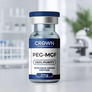
PEG-MGF Research Peptide
HPLC and MS verified · certificate with every batch · pricing announced when stock arrives
PEG-MGF vs MGF: The Real Difference
This is the question most people searching for PEG-MGF want answered, so here is the comparison in plain terms.
How long it lasts
Native MGF is a short, unprotected peptide that enzymes can break down quickly. Pegylation is designed to slow both enzyme breakdown and kidney clearance, so PEG-MGF is expected to last far longer. How much longer has not been measured in a published study, and it would depend heavily on the size and position of the PEG chain.
Where it acts
Natural MGF appears to work as a local, autocrine and paracrine signal, made and used within the tissue under strain (PMID: 12692175). A longer-lasting molecule has more time to travel. So the most interesting research question about PEG-MGF is whether making MGF last longer changes it from a local signal into a body-wide one, and what that would mean for its effects.
How strongly it acts at its target
Pegylation can reduce how strongly a molecule acts at its target, as the pegylated IGF-1 work shows (PMID: 29634992). MGF’s own target is still debated, so it is not yet known how a PEG chain would affect it. That makes PEG-MGF a genuinely open research question rather than a known quantity.
What carries over from MGF research
Everything that makes MGF interesting also applies to PEG-MGF as a starting point: the stem cell findings, the heart and nerve studies, and the debate about how the synthetic peptide compares with the natural one. A 2014 study from two pharmaceutical companies could not reproduce several classic cell findings with the MGF peptide (PMID: 24253050), and a 2016 study found that the 24-residue MGF peptide did not activate the IGF-1 receptor, while full-length MGF protein did (PMID: 26991004). Any PEG-MGF study would build on that foundation, and its results would help settle some of the same questions.
Compared with other ways of making peptides last
Pegylation is not the only way to extend a peptide’s life. CJC-1295 (With DAC) uses a different trick, a reactive group that lets the peptide bind to albumin in the blood. IGF-1 LR3 takes another route, changing the amino acid sequence so the molecule escapes the proteins that normally bind IGF-1. Our article on CJC-1295 with DAC vs without DAC shows how a single half-life extension can change a peptide’s whole research profile, which is exactly the question PEG-MGF raises.
The Questions a PEG-MGF Study Would Answer
PEG-MGF sits at an exciting intersection: a well-studied modification applied to a fascinating growth signal. Here are the questions researchers would be looking to answer.
The half-life. How long does PEG-MGF actually last, and how does that change with the size of the PEG chain? The pegfilgrastim and pegylated IGF-1 data suggest the effect could be dramatic.
The activity. Does PEG-MGF keep the cell effects reported for native MGF, such as satellite cell activity and protection from cell death, or does the PEG chain dampen them? Side-by-side cell experiments with native and pegylated peptide would answer that quickly.
Local or systemic. Does making MGF last longer change where it acts? Tracking a labelled peptide through the body would show whether it stays near where it is delivered or spreads widely.
The design. Where should the PEG be attached, and should it be releasable? The protease-sensitive linker work on IGF-1 points to designs that could last in the blood and release a fully active peptide where tissue-damage enzymes are present, so far shown only in laboratory tests (PMID: 29634992).
The immune response to PEG itself. A study of samples from the general population detected antibodies against PEG in about 72% of contemporary samples, mostly at low levels (PMID: 27804292), and in a study of pegylated asparaginase, anti-PEG antibodies were closely associated with rapid clearance of the drug (PMID: 17516438). Researchers working with any pegylated molecule now factor this in, and it is an interesting variable in animal studies too.
The Technical Breakdown: PEG Chemistry, Chain Size and Structure
This section is for readers who want the molecular detail. Everything above makes sense without it.
What PEG is
Polyethylene glycol is a chain of repeating ethylene oxide units, written as -(CH2-CH2-O)n-. For pegylation it is usually used as methoxy-PEG, capped at one end so it attaches at the other. PEG reagents are described by average molecular weight, such as 2 kDa, 5 kDa, 20 kDa or 40 kDa, and come in linear or branched forms (PMID: 26869412).
Where PEG can attach to MGF
The native MGF sequence is YQPPSTNKNTKSQRRKGSTFEERK. Common pegylation chemistries target free amine groups, which on MGF means the N-terminus and the side chains of its three lysines. Attaching at different positions produces different molecules, and attaching more than one PEG chain produces a mixture. Site-specific methods, such as the N-terminal approach used for pegfilgrastim (PMID: 15078138), give a more uniform product.
Why there is no single “PEG-MGF” structure
Because of those choices, PEG-MGF is better thought of as a family of possible molecules than a single defined compound. The PEG size, the number of PEG chains, the attachment site and the version of the MGF peptide underneath can all vary. MGF itself comes in several versions, including the native peptide, a C-terminal amide (PMID: 22328223) and a variant with histidine at position 23 (PMID: 28035768). Supplier listings for PEG-MGF do not agree on its molecular weight for exactly this reason. For research, that makes a clear description of the exact structure essential.
Why a pegylated peptide gives a spread of masses
PEG reagents are polymers, so each batch contains chains of slightly different lengths. In mass spectrometry, a pegylated peptide therefore appears as a distribution of peaks spaced 44 Da apart, the mass of one ethylene oxide unit, rather than a single sharp peak. Reading that distribution confirms both the peptide and the average PEG size.
Why Identity Testing Matters Even More for a Pegylated Peptide
With most peptides, a certificate of analysis answers two questions: is it the right molecule, and how pure is it? With a pegylated peptide, there are more. Which MGF peptide is underneath? How big is the PEG chain? How many chains are attached? Is any unpegylated peptide left over? For a compound without a single agreed structure, those answers are what make research results meaningful and repeatable.
That is the standard we apply across our range, and it is the standard PEG-MGF is held to.
What every Crown Peptides batch comes with
Every batch we sell is tested before it goes on sale. HPLC (high-performance liquid chromatography) separates the target compound from anything else in the vial and measures its purity. Mass spectrometry confirms identity by measuring the molecule’s mass. Results are published as a certificate of analysis for that exact batch on our Lab Reports page, with the batch number printed on the vial you receive. Our guides on how to read a peptide certificate of analysis and how to tell if peptides are real explain what to look for.
Stored, packed and dispatched properly
Our compounds are supplied as lyophilised (freeze-dried) powder, the most stable form for storage and transport. Vials are kept cold, sealed and away from light and moisture, as described on our storage information page. Weekday orders placed before 2pm are dispatched the same day from the UK, and UK delivery is free on orders over £100.
A UK company you can actually talk to
Crown Peptides is a UK company. Our team replies within 24 hours on business days, and WhatsApp is the quickest way to reach us. If you have a question about a certificate, a batch or a compound in our range, you can get in touch.
Related Compounds Worth Knowing About
PEG-MGF sits where growth factor research meets half-life engineering. These compounds explore the same IGF-1 system or the same idea of making a peptide last, and most sit in our musculoskeletal research peptides category.
MGF
Native MGF is the unmodified parent peptide: short-lived, local and the subject of three decades of research. Start with our MGF peptide guide for the full story.
IGF-1 LR3
IGF-1 LR3 is a long-acting analogue of mature IGF-1, made to last by changing its sequence rather than adding PEG. It is the go-to compound for studying the IGF-1 receptor directly. Our IGF-1 LR3 guide explains how it was designed.
CJC-1295 (With DAC)
CJC-1295 (With DAC) is a growth hormone-releasing hormone analogue with a drug affinity complex that binds albumin, one of the best-known examples of half-life extension in peptide research. It is the natural comparison for anyone interested in what a longer-lasting version of a short peptide can do.
Tesamorelin
Tesamorelin is another growth hormone-releasing hormone analogue, stabilised with a small chemical group at its N-terminus. It sits at the top of the growth hormone and IGF-1 chain that MGF branches off at the end.
BPC-157 and TB-500
BPC-157 and TB-500 (Thymosin Beta-4) are two of the most studied tissue repair peptides. They come from different biology from MGF but are often studied in the same muscle, tendon and recovery models.
For a wider view, see our guide to the best peptides for muscle research, or our explainer on peptide half-life.
Common Questions About PEG-MGF
What is PEG-MGF?
PEG-MGF is the mechano growth factor peptide with polyethylene glycol chains attached. Pegylation is designed to slow breakdown and kidney clearance so the peptide lasts longer than native MGF.
What is the difference between PEG-MGF and MGF?
MGF is a short, unprotected 24-amino acid peptide that is broken down quickly and is thought to act locally. PEG-MGF is the same peptide with PEG attached, which is designed to make it last much longer and may change where it acts in the body.
Has PEG-MGF been studied?
PubMed lists no peer-reviewed study of PEG-MGF itself. The research context comes from studies of the MGF peptide and from decades of work on pegylated proteins, including a human study of pegylated IGF-1.
How long does PEG-MGF last?
Its half-life has not been measured in a published study. Pegylation extended the half-life of filgrastim from 3.5 to 3.8 hours to 42 hours, and pegylated IGF-1 reached 140 to 200 hours in people, which shows the scale of change pegylation can produce in related proteins.
Is PEG-MGF the same as IGF-1?
No. PEG-MGF is built on the MGF peptide, which comes from the tail of the IGF-1Ec precursor and contains none of the IGF-1 molecule itself.
Why do PEG-MGF molecular weights vary between sources?
Because there is no single standard structure. The size of the PEG chain, the number of chains and where they attach all change the final mass. That is why a certificate showing the exact identity of each batch matters so much.
Is PEG-MGF legal in the UK?
PEG-MGF is not listed under the Misuse of Drugs Act, and research compounds can be bought in the UK for laboratory research. Mechano growth factors do appear on the World Anti-Doping Agency’s prohibited list, which applies to competitive sport. Our article are peptides legal in the UK explains the wider legal picture.
Can I buy PEG-MGF in the UK?
Yes. Crown Peptides supplies PEG-MGF in 2mg vials, within our musculoskeletal research peptides category. With a pegylated peptide, the certificate matters even more than usual, because it is the only way to see which peptide and which PEG size you actually have.
How should pegylated peptides be stored?
Like other research peptides, they are best kept as a lyophilised powder, cold, sealed and away from light and moisture. Our storage information page covers the details.
Read the certificate before you order
Every batch is published openly — identity by mass spectrometry, purity by HPLC, and the batch number printed on the vial you receive.
Open the COA libraryA Short Signal, Given a Long Life
PEG-MGF brings together two very different ideas. One is a growth signal that muscle seems to make for a moment, right where it is needed. The other is one of the most successful tools in pharmaceutical chemistry, a polymer that can turn a molecule lasting hours into one lasting days.
The research on pegylated IGF-1 shows that this kind of change does more than extend a timeline. It can change how the body responds to a growth factor. That is what makes PEG-MGF such an interesting research question. Whether a longer-lasting MGF keeps its local character, gains new effects or behaves in ways nobody has predicted yet is exactly the kind of thing well-designed laboratory work can find out.
To explore the compounds behind this story, see our PEG-MGF Research Peptide page or browse our musculoskeletal research peptides, every batch HPLC and MS verified, with its certificate published in full and dispatched from the UK.
References
- Hill M, Goldspink G. Expression and splicing of the insulin-like growth factor gene in rodent muscle is associated with muscle satellite (stem) cell activation following local tissue damage. J Physiol. 2003;549:409-18. PMID: 12692175
- Dominikowski A, Rękoś Z, Olejarz M, et al. The emerging landscape of performance-enhancing peptides modulating GH-IGF1 axis: bridging the gap between clinical evidence and patient self-administration. Front Endocrinol (Lausanne). 2026;17:1822475. PMID: 42395176
- Abuchowski A, McCoy JR, Palczuk NC, et al. Effect of covalent attachment of polyethylene glycol on immunogenicity and circulating life of bovine liver catalase. J Biol Chem. 1977;252:3582-6. PMID: 16907
- Harris JM, Chess RB. Effect of pegylation on pharmaceuticals. Nat Rev Drug Discov. 2003;2:214-21. PMID: 12612647
- Turecek PL, Bossard MJ, Schoetens F, et al. PEGylation of Biopharmaceuticals: A Review of Chemistry and Nonclinical Safety Information of Approved Drugs. J Pharm Sci. 2016;105:460-475. PMID: 26869412
- Molineux G. The design and development of pegfilgrastim (PEG-rmetHuG-CSF, Neulasta). Curr Pharm Des. 2004;10:1235-44. PMID: 15078138
- Yamaoka T, Tabata Y, Ikada Y. Distribution and tissue uptake of poly(ethylene glycol) with different molecular weights after intravenous administration to mice. J Pharm Sci. 1994;83:601-6. PMID: 8046623
- Braun AC, Gutmann M, Mueller TD, et al. Bioresponsive release of insulin-like growth factor-I from its PEGylated conjugate. J Control Release. 2018;279:17-28. PMID: 29634992
- Kletzl H, Guenther A, Höflich A, et al. First-in-man study with a novel PEGylated recombinant human insulin-like growth factor-I. Growth Horm IGF Res. 2017;33:9-16. PMID: 28110155
- Sama DM, Carlson SW, Joseph B, et al. Assessment of systemic administration of PEGylated IGF-1 in a mouse model of traumatic brain injury. Restor Neurol Neurosci. 2018;36:559-569. PMID: 29889090
- Doroudian G, Pinney J, Ayala P, et al. Sustained delivery of MGF peptide from microrods attracts stem cells and reduces apoptosis of myocytes. Biomed Microdevices. 2014;16:705-15. PMID: 24908137
- Peña JR, Pinney JR, Ayala P, et al. Localized delivery of mechano-growth factor E-domain peptide via polymeric microstructures improves cardiac function following myocardial infarction. Biomaterials. 2015;46:26-34. PMID: 25678113
- Fornaro M, Hinken AC, Needle S, et al. Mechano-growth factor peptide, the COOH terminus of unprocessed insulin-like growth factor 1, has no apparent effect on myoblasts or primary muscle stem cells. Am J Physiol Endocrinol Metab. 2014;306:E150-6. PMID: 24253050
- Janssen JA, Hofland LJ, Strasburger CJ, et al. Potency of Full-Length MGF to Induce Maximal Activation of the IGF-I R Is Similar to Recombinant Human IGF-I at High Equimolar Concentrations. PLoS One. 2016;11:e0150453. PMID: 26991004
- Yang Q, Jacobs TM, McCallen JD, et al. Analysis of Pre-existing IgG and IgM Antibodies against Polyethylene Glycol (PEG) in the General Population. Anal Chem. 2016;88:11804-11812. PMID: 27804292
- Armstrong JK, Hempel G, Koling S, et al. Antibody against poly(ethylene glycol) adversely affects PEG-asparaginase therapy in acute lymphoblastic leukemia patients. Cancer. 2007;110:103-11. PMID: 17516438
- Esposito S, Deventer K, Van Eenoo P. Characterization and identification of a C-terminal amidated mechano growth factor (MGF) analogue in black market products. Rapid Commun Mass Spectrom. 2012;26:686-92. PMID: 22328223
- Cox HD, Miller GD, Eichner D. Detection and in vitro metabolism of the confiscated peptides BPC 157 and MGF R23H. Drug Test Anal. 2017;9:1490-1498. PMID: 28035768
Working with PEG-MGF Research Peptide
Coming soon to Crown Peptides. Every batch will be HPLC and MS tested, with its certificate published before it goes on sale.
See PEG-MGF Research Peptide