

Most research peptides earn their name from one specific, well-defined job. VIP earns its name from the very first job researchers happened to discover — vasodilation in the gut — and then spent the following decades finding that same compound doing something functionally important in nearly every other major organ system in the body.
Vasoactive Intestinal Peptide, VIP for short, has one of the broadest documented physiological footprints of any research peptide discussed in Crown Peptides' catalogue — touching the immune system, the central nervous system, the gut, the lungs, and the body's own internal circadian clock, all through a shared receptor family with remarkably wide tissue distribution.
VIP is sold by Crown Peptides for laboratory research use only and is not approved for human consumption.
The short version
- Twenty-eight residues, acting at the VPAC1 and VPAC2 receptors.
- Found across gut, lung, brain and immune tissue — unusually widespread.
- Originally isolated from intestine, hence the name.
- Breadth is the difficulty: the literature spans several unrelated fields.
A 28-Amino-Acid Peptide From the Secretin Family
VIP is a 28-amino-acid peptide, originally isolated from and named for its vasodilating effects within intestinal tissue, though that original characterisation turned out to represent only a fraction of its actual physiological role. Structurally, VIP belongs to the secretin family of peptide hormones, a related group of signalling molecules that includes secretin, glucagon, and pituitary adenylate cyclase-activating polypeptide (PACAP) — VIP's closest structural relative, sharing considerable sequence homology and, notably, some overlapping receptor activity.
VIP is secreted by the enteric nervous system — the extensive, semi-independent network of neurons embedded within the gut wall, sometimes described as the body's "second brain" for its capacity to regulate digestive function with a considerable degree of autonomy from the central nervous system. That enteric origin is consistent with VIP's earliest documented effects, but VIP-producing neurons and VIP receptors are, in fact, distributed across a remarkably wide range of tissues well beyond the digestive system.
PACAP: The Close Structural Relative Worth Knowing
VIP's relationship to PACAP deserves explanation, since the two peptides are so frequently discussed together in the research literature. PACAP and VIP share a common evolutionary origin and considerable sequence similarity, and critically, PACAP is capable of activating the same VPAC1 and VPAC2 receptors that VIP engages, alongside a third receptor, PAC1, for which PACAP shows considerably higher affinity than VIP does.
That receptor overlap means research studying either peptide in isolation needs to account for the possibility of shared downstream effects, and it's part of why researchers frequently design comparative studies examining both peptides together — distinguishing which specific physiological effects are genuinely VIP-selective, mediated predominantly through VPAC receptor engagement, versus which effects might be better attributed to the PAC1-mediated activity more specific to PACAP. That receptor-selectivity question remains an active and genuinely important consideration across this entire peptide family's research literature.
- Classification
- Vasoactive intestinal peptide, a 28-amino-acid neuropeptide
- CAS Number
- 40077-57-4
- Molecular Weight
- 3325.79 Da
- Molecular Formula
- C147H238N44O42S
- Sequence
- His-Ser-Asp-Ala-Val-Phe-Thr-Asp-Asn-Tyr-Thr-Arg-Leu-Arg-Lys-Gln-Met-Ala-Val-Lys-Lys-Tyr-Leu-Asn-Ser-Ile-Leu-Asn-NH2
Two Receptors, Two Distinct Vasodilation Mechanisms
VIP's biological actions are mediated primarily through two G-protein coupled receptors, VPAC1 and VPAC2, both members of the Class B secretin receptor family, alongside some activity at a third receptor, NPR-C. Research examining VIP-induced vasodilation specifically has documented that these receptors work through genuinely distinct mechanisms depending on their location: the VPAC1 receptor, positioned on the vascular endothelium, produces vasodilation by generating nitric oxide (NO) — the same signalling molecule central to much of the body's vascular tone regulation generally.
The VPAC2 receptor, by contrast, is expressed predominantly in the outer smooth muscle layers of blood vessels and produces vasodilation independently of nitric oxide, in response to VIP released directly from intramural nerves within the vessel wall itself. That mechanistic split — one receptor working through the classic nitric oxide pathway, the other through an entirely separate, NO-independent route, both converging on the same functional outcome — is a genuinely elegant piece of physiological redundancy, and it illustrates the broader pattern of layered, multi-mechanism signalling that recurs throughout VIP's research literature.
Class B GPCRs: A Distinctive Receptor Architecture
It's worth understanding why VPAC1 and VPAC2's classification as Class B G-protein coupled receptors matters mechanistically. Class B GPCRs, sometimes called secretin-family receptors, are structurally distinct from the more common Class A GPCR family, which includes the receptors targeted by many conventional small-molecule pharmaceuticals. Class B receptors typically feature a considerably larger extracellular domain, which plays a central role in initially capturing and binding the peptide ligand before that ligand engages the receptor's transmembrane core.
That structural distinction has practical research implications: compounds targeting Class B receptors, including VIP itself, are generally peptides rather than small molecules, given how the large extracellular binding domain is typically better suited to engaging a peptide-sized ligand than a considerably smaller small-molecule compound. Understanding this receptor architecture helps explain why VIP's activity, along with that of its structural relatives secretin, glucagon, and PACAP, has historically been harder to replicate with conventional small-molecule drug design approaches, reinforcing the research value of studying these peptides directly.
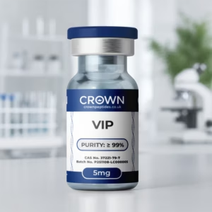
VIP Research Peptide
HPLC and MS verified · batch certificate published · dispatched from the UK before 2pm
The Suprachiasmatic Nucleus: VIP as a Circadian Synchroniser
One of VIP's most mechanistically well-characterised research roles concerns the circadian clock itself. VIP is released by retinorecipient cells within the suprachiasmatic nucleus (SCN) — the small hypothalamic structure widely recognised as the body's master circadian pacemaker, directly receiving light input from the retina and using that input to synchronise the body's internal clock to the external day-night cycle.
Research published in Nature Communications has documented that VIP controls the broader suprachiasmatic circadian clock network specifically through ERK1/2 signalling and its negative regulator DUSP4 — a defined molecular pathway responsible for what researchers describe as VIP-directed circadian reprogramming. Functionally, VIP acts as a synchronising agent among individual SCN neurons, helping maintain coherent, coordinated oscillatory activity across the broader neuronal network responsible for generating the body's roughly 24-hour internal rhythm, rather than each neuron oscillating independently and asynchronously.
Retinorecipient Cells: The Light-to-Clock Connection
The retinorecipient cell population responsible for VIP release within the SCN deserves a closer look, since it represents the specific anatomical bridge connecting external light exposure to internal circadian timing. These cells receive direct input from the retinohypothalamic tract, a dedicated neural pathway carrying light information from specialised retinal cells straight to the SCN, bypassing the visual cortex and conscious visual processing entirely — reflecting how fundamental and evolutionarily ancient the light-to-clock signalling pathway is.
That direct anatomical connection is precisely why VIP release from these specific SCN cell populations can function as an effective real-time synchronising signal: it sits at the exact junction where external light information is converted into an internal neurochemical signal capable of resetting and coordinating the master clock's cellular network, making VIP research a genuinely central piece of understanding how environmental light exposure translates into coherent, network-wide circadian timing at the cellular level.
Why Synchronisation Matters More Than Individual Timekeeping
It's worth explaining why this synchronising role is so significant within circadian research specifically. Individual SCN neurons are each capable of generating their own independent oscillatory rhythm through cell-autonomous molecular clock machinery, but a functional, coherent circadian output requires those thousands of individual neuronal oscillators to remain synchronised with one another — without that synchronisation, the SCN's output signal would become desynchronised and functionally incoherent, even if each individual neuron's internal clock remained technically intact.
VIP's research role addresses precisely that synchronisation requirement, making it a genuinely distinct research target from compounds addressing circadian biology through other mechanisms, such as melatonin-focused research or Epitalon's pineal gland-centred mechanism discussed elsewhere in Crown Peptides' catalogue. Where those compounds work more directly on the downstream hormonal output of circadian timing, VIP's research addresses the upstream network coordination problem that determines how coherently the master clock itself functions in the first place.
ERK1/2 and DUSP4: Understanding the Molecular Switch
The ERK1/2 and DUSP4 signalling pathway underlying VIP's circadian synchronising role deserves further explanation for researchers less familiar with these specific molecules. ERK1/2 (extracellular signal-regulated kinases) are part of the broader MAPK signalling family, a well-characterised intracellular signalling cascade involved in translating external receptor signals into changes in gene expression and cellular behaviour, relevant across an enormous range of physiological contexts well beyond circadian biology alone.
DUSP4 functions as a negative regulator within this pathway, dephosphorylating and thereby deactivating ERK1/2 once its signal has been appropriately transmitted — a built-in braking mechanism ensuring the signalling cascade doesn't run unchecked. Research identifying this specific ERK1/2-DUSP4 regulatory loop as central to VIP's circadian synchronising function represents a genuinely detailed, molecularly precise mechanistic finding, offering researchers specific, well-defined molecular targets for further dissecting exactly how VIP achieves its network-wide synchronising effect at the cellular level.
Immunoregulatory Research: A Broad Anti-Inflammatory Profile
VIP's immunoregulatory research profile is similarly broad. Research has documented anti-inflammatory effects across multiple immune cell populations, including T cells, macrophages, and plasmacytoid dendritic cells — a genuinely wide range of distinct immune cell types, each playing different roles within the broader immune response, all shown to be influenced by VIP signalling in documented research.
Mechanistically, VIP's engagement with VPAC receptors initiates cAMP-dependent transcriptional cascades, and research suggests this signalling may recalibrate inflammatory tone alongside several other physiological variables simultaneously — neural plasticity, endocrine rhythms, and vascular dynamics among them. That breadth reflects the same underlying pattern seen throughout VIP's research literature: a single receptor-ligand system, engaged across genuinely diverse tissue contexts, producing correspondingly diverse downstream physiological consequences.
cAMP Signalling: The Common Downstream Thread
It's worth highlighting cAMP's recurring role across VIP's mechanistic profile, since it represents a genuine unifying thread running through the compound's otherwise diverse tissue-specific effects. Cyclic AMP (cAMP) is one of the most widely used second messenger molecules in cell signalling generally, translating an initial receptor-binding event at the cell surface into a broad range of downstream intracellular consequences, frequently including changes in gene transcription, enzyme activity, and cellular metabolism.
Because both VPAC1 and VPAC2 signal predominantly through cAMP-generating pathways, VIP's remarkably diverse tissue-specific effects — vasodilation, immune modulation, circadian synchronisation, smooth muscle relaxation — can be understood as different tissue-specific interpretations of essentially the same core second-messenger signal, shaped by which particular downstream cAMP-responsive genes and processes happen to be active within each specific cell type and tissue context. That shared signalling architecture is a useful unifying lens for researchers trying to make sense of VIP's otherwise sprawling physiological reach.
The Gut-Immune Connection
Given VIP's enteric nervous system origins, its research profile within gut immunology deserves particular attention. Research has examined VIP's interaction with enteric glial networks — the gut's own specialised support cells, analogous in some respects to glial cells within the central nervous system — alongside the substantial population of immune cells residing within the gut microenvironment specifically. That research positions VIP as a genuine coordinating signal at the intersection of the enteric nervous system and gut-resident immune populations, consistent with the gut's well-established role as one of the body's largest and most immunologically active organs.
This gut-immune research thread connects directly back to VIP's broader anti-inflammatory profile discussed above, offering researchers a tissue-specific research context — the gastrointestinal system — in which to study VIP's immunoregulatory mechanisms in particularly fine mechanistic detail, given how extensively gut immunology and enteric nervous system biology have already been characterised independently in the broader research literature.
Fetal Development and Broader Reproductive Research
VIP's research footprint extends even into developmental biology, with documented roles in fetal development processes — a research thread reflecting VIP's broad expression pattern across developing tissue well before birth. That developmental relevance adds yet another dimension to VIP's already extensive research profile, though it remains a considerably less extensively characterised area compared with the vasodilation, circadian, and immune research discussed at length throughout this piece.
This developmental research thread is worth mentioning specifically because it reinforces the broader pattern evident throughout VIP's research history: a signalling molecule this widely expressed, across tissue types spanning from early fetal development through to the fully mature adult nervous, immune, and vascular systems, tends to accumulate research relevance across an unusually broad range of physiological contexts over time, simply as a consequence of how extensively distributed its receptor expression turns out to be.
Pulmonary Research and Smooth Muscle Relaxation
VIP's research relevance extends into pulmonary physiology as well, given its documented effects on smooth muscle relaxation more broadly — a mechanism directly relevant to airway tone regulation in lung tissue, alongside its vascular smooth muscle effects discussed earlier. That pulmonary research thread connects VIP to broader research questions around bronchial smooth muscle regulation and airway inflammation, given the same VPAC receptor-mediated mechanisms documented in vascular tissue operate similarly within pulmonary smooth muscle.
Endocrine Secretion Research: Beyond the Gut
VIP's documented influence on exocrine and endocrine secretions extends its research relevance into hormonal regulation more broadly, beyond the gut-specific secretory functions its name originally referenced. Research has examined VPAC receptor expression across several pituitary-derived cell lines specifically, suggesting a role for VIP in regulating the synthesis and release of various pituitary hormones — connecting VIP's research profile to the broader neuroendocrine signalling territory discussed extensively elsewhere in Crown Peptides' catalogue, including the GHRH-pathway and GH-secretagogue peptides.
That pituitary-adjacent research thread adds a further dimension to VIP's already broad physiological reach, reinforcing the same fundamental theme running throughout this compound's research literature: wherever VPAC receptors are expressed, VIP appears capable of exerting a measurable, mechanistically documented physiological influence, and pituitary tissue turns out to be yet another site where that receptor expression, and the corresponding research relevance, has been confirmed.
Dosage in Research Settings
VIP's research base is concentrated in cellular, tissue, and animal model studies rather than standardised human systemic dosing trials. In suprachiasmatic nucleus research specifically, concentrations between roughly 150 nM and 10 uM have been used to characterise its dose-dependent effect on circadian clock-neuron synchrony, with both the degree and duration of the resulting desynchronisation depending on dose and timing. Those figures describe laboratory tissue-culture concentrations, not a human administration guideline, and no completed human dose-ranging trials establish an equivalent systemic figure.
Reported Benefits in the Research Data
VIP's research record spans two major domains: circadian biology, where it's considered a key candidate molecule synchronising clock gene expression in the suprachiasmatic nucleus and is critical for circadian regulation of glucocorticoids via the HPA axis, and immunology, where a major pharmacological review characterised it as playing central roles in immune cellular and molecular events, including generating regulatory T cells and tolerogenic dendritic cells relevant to inflammatory and autoimmune disease research.
Side Effects Reported in Studies
Because VIP's evidence base sits predominantly in cellular and animal research rather than completed human systemic trials, a human safety profile in the sense of a controlled clinical dataset doesn't exist for it in the peer-reviewed literature. Its dose-dependent effects on neuronal synchrony in circadian research specifically illustrate why concentration and timing both matter for how VIP behaves in any given experimental context, rather than the compound having a single fixed effect regardless of dose.
Manufactured to Research Standard
VIP's 28-amino-acid structure, considerably larger than many shorter research peptides, requires particularly careful synthesis and purity verification to ensure reliable, reproducible research results.
Crown Peptides tests every batch of VIP for HPLC purity and mass spectrometry identity confirmation, with a batch-specific certificate of analysis provided for every order.
Storage follows the standard protocol used across the peptide range: supplied as a lyophilised powder, stable when kept cold and shielded from light and moisture, with reconstituted solution requiring refrigeration and use within the recommended window.
Trigeminal and Cerebrovascular Research
VIP's research profile also extends into the trigeminal ganglion and cerebrovascular tissue, where immunohistochemical research has documented localisation of VIP, its close relative PACAP, and their shared receptors within basal brain blood vessels and the trigeminal nervous system specifically. That anatomical localisation research offers relevant context for researchers studying vascular headache and migraine-adjacent physiology, given how centrally the trigeminovascular system — the network connecting trigeminal sensory neurons to cerebral blood vessels — features in that broader area of neuroscience research.
That cerebrovascular research thread ties back neatly to VIP's fundamental vasodilating mechanism discussed at the outset of this piece, demonstrating that even VIP's most "classic," longest-documented mechanism continues to generate genuinely new and clinically relevant research questions when examined within specific, well-characterised anatomical contexts like the cerebrovascular and trigeminal systems.
VIP Compared With PACAP and Other Related Peptides
VIP's closest structural and functional relative, PACAP, shares considerable receptor overlap and physiological reach, and the two peptides are frequently studied together given their related signalling architecture and overlapping VPAC receptor engagement. Within Crown Peptides' broader catalogue, VIP's circadian research role also invites comparison with Epitalon, discussed extensively elsewhere, given both compounds' research relevance to circadian and pineal-adjacent biology, even though their specific mechanisms — network synchronisation for VIP, melatonin-pathway restoration for Epitalon — are genuinely distinct.
- Structure: 28-amino-acid peptide, member of the secretin peptide family alongside secretin and PACAP.
- Receptors: VPAC1 and VPAC2 (Class B GPCRs), plus some activity at NPR-C.
- Circadian role: synchronises suprachiasmatic nucleus neurons via ERK1/2 and DUSP4 signalling.
- Immune research: documented anti-inflammatory effects on T cells, macrophages, and dendritic cells.
- Vasodilation mechanism: VPAC1 via nitric oxide (endothelium); VPAC2 independently of nitric oxide (smooth muscle).
Frequently Asked Questions:
What is VIP?
A 28-amino-acid neuropeptide, originally identified for its vasodilating effects in intestinal tissue, now studied for a far broader range of effects spanning the immune system, circadian biology, the gut, and the lungs.
What is VIP Peptide Used For?
VIP (Vasoactive Intestinal Peptide) is used primarily in research to study its neuroprotective, anti-inflammatory, and immune-modulating effects, notably in protocols addressing chronic inflammatory response syndromes (CIRS) and biotoxin illnesses. It acts as a signaling neuropeptide that helps regulate blood flow, smooth muscle relaxation, and immune system balance throughout the body.
How do VIP's two receptors differ in producing vasodilation?
VPAC1, located on the vascular endothelium, produces vasodilation by generating nitric oxide. VPAC2, located in vascular smooth muscle, produces vasodilation independently of nitric oxide, in response to VIP released from intramural nerves.What role does VIP play in circadian rhythm research?
VIP synchronises individual suprachiasmatic nucleus neurons with one another, maintaining coherent network-wide oscillatory activity, via ERK1/2 and DUSP4 signalling — addressing network coordination rather than each neuron's individual timekeeping.
What immune cell types has VIP research examined?
Research has documented anti-inflammatory effects across T cells, macrophages, and plasmacytoid dendritic cells, mediated through cAMP-dependent transcriptional cascades downstream of VPAC receptor engagement.
How is VIP related to PACAP?
PACAP shares considerable sequence homology and activates the same VPAC1/VPAC2 receptors, alongside a third receptor, PAC1, for which it shows considerably higher affinity than VIP.
Cancer Research: An Emerging and Complex Angle
VIP's receptor system has also drawn research interest within oncology, though the findings here are genuinely more complex and, in some respects, seemingly contradictory compared with VIP's more straightforwardly beneficial research profile elsewhere. Research has identified VPAC2 receptor signalling as promoting pancreatic cancer cell growth in some models, while separately decreasing the immunogenicity of the tumour microenvironment — findings that position VPAC2 as a potential therapeutic target for antagonism in this specific cancer context, the opposite research direction from VIP's broadly anti-inflammatory role documented elsewhere.
Separately, researchers have identified and characterised VIP receptor antagonists — compounds that block rather than activate VIP receptor signalling — specifically for their anti-leukemia activity in preclinical research models. That dual research direction, with VIP receptor activation studied for broadly anti-inflammatory purposes in most contexts while VIP receptor blockade is separately studied for anti-cancer purposes in specific tumour contexts, illustrates a genuinely important principle worth understanding: a signalling pathway's research value can point in opposite therapeutic directions depending on the specific tissue and disease context under investigation, and VIP's oncology-adjacent literature is a clear illustration of that context-dependent complexity.
Read the certificate before you order
Every batch is published openly — identity by mass spectrometry, purity by HPLC, and the batch number printed on the vial you receive.
Open the COA libraryThe Bottom Line
VIP's research profile is defined by breadth rather than narrow specialisation: a single peptide, signalling through a shared receptor family expressed across the gut, the vasculature, the immune system, and the brain's own master circadian clock. That physiological reach, documented across genuinely distinct organ systems through mechanistically well-characterised pathways, is precisely what keeps VIP such an actively studied compound across such a wide swath of contemporary physiological and immunological research.
References
- Delgado M, Pozo D, Ganea D. The significance of vasoactive intestinal peptide in immunomodulation. Pharmacol Rev. 2004;56(2):249-290. https://pubmed.ncbi.nlm.nih.gov/15169929/
Working with VIP Research Peptide
Batch-tested material, published certificates, and same-day dispatch on orders placed before 2pm.
See VIP Research Peptide